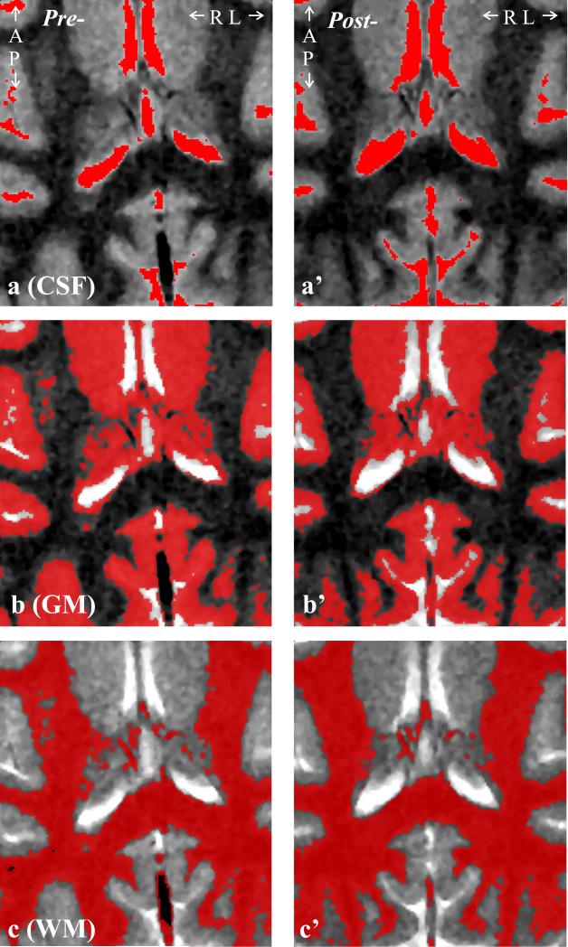
Fig. 2.
Left: Axial T2-weighted TSE image shows the anatomical coverage of the 4.0×3.5 cm2 VOI, overlaid with the: a: CSF, b: GM and c: WM masks generated by the FireVoxel package. Note the tissue type differentiation performance.
Right: a’-c’: Same as a-c, post-infection of the same female macaque.

