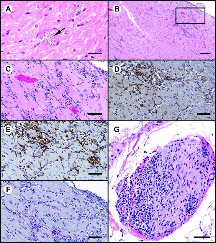
Figure 3.
Sections of myocardium demonstrating areas of inflammation and characterization of the inflammatory cell infiltrates by IHC. (A) Cluster of T. cruzi amastigotes (arrow). Hematoxylin and eosin stain; bar, 20 μm. (B) Low-power magnification demonstrating moderate to marked subepicardial inflammatory cell infiltrates. Hematoxylin and eosin stain; bar 300μm. The boxed area is shown below. (C) High-power magnification from section in panel B. Inflammatory cell infiltrates are primarily mononuclear cells with fewer neutrophils. Hematoxylin and eosin stain; bar, 100 μm. (D and E) IHC peroxidase procedure with diaminobenzidine chromagen (brown). A considerable proportion of the mononuclear cell infiltrates are positive for (D) CD8 (lymphoid origin) and (E) CD68 (monocyte–macrophage origin). Bar, 100 μm. (F) IHC alkaline phosphatase procedure with Vector red chromagen (red). A small proportion of inflammatory cells are CD4+. Bar, 100 μm. G) Section of nerve at the heart base that is infiltrated by inflammatory cells. Hematoxylin and eosin stain; bar, 80 μm.

