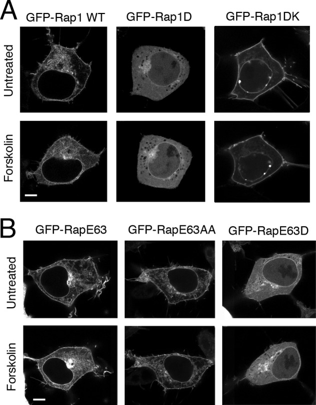
FIGURE 6.

Localization of Rap1 mutants in cells. A, the introduction of basic amino acids restores plasma membrane relocalization of the Rap1D mutant. Micrographs of HEK293 cells transfected with GFP-tagged Rap1 constructs, GFP-Rap1WT, GFP-Rap1D, and GFP-Rap1DK, are shown. Images were taken before and 20 min after forskolin treatment. For each transfection, a representative cell is shown. The scale bar is 5 μm in length. B, localization of RapE63 is regulated by PKA phosphorylation. Shown here are micrographs of HEK293 cells transfected with the GFP-tagged RapE63 constructs, GFP-RapE63 WT, GFP-RapE63AA, and GFP-RapE63D. For each transfection, a representative cell is shown before and after forskolin treatment. The scale bar is 5 μm in length.
