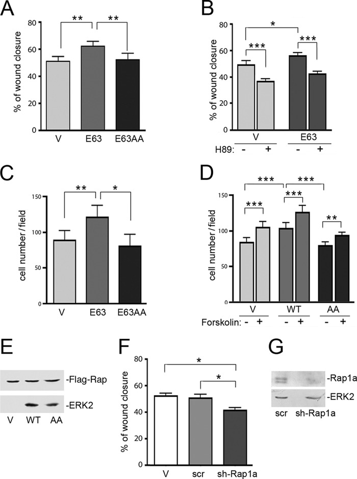
FIGURE 8.
Cell migration assays show Rap1-dependent migration. A, wound healing assay. Stably transfected H1299 cells expressing pcDNA3 vector (V), FLAG-RapE63 (E63), or FLAG-RapE63AA (E63AA) were examined by a wound healing assay as described under “Experimental Procedures.” The bar graph shows the percentage of wound closure across the scratch at 8 h. The values shown are the average of 5 independent experiments, ± S.E. **, denotes statistical significance at <0.01. B, wound healing assay. Stably transfected H1299 cells expressing pcDNA vector (V) or FLAG-RapE63 (E63) were prepared for a wound healing assay as described under “Experimental Procedures.” Then cells were incubated in 1% serum with (+) or without (−) H89. Cell migration across the gap was monitored and analyzed after 8 h. The bar graph shows the percentage of wound closure. n = 3, ± S.E. *, denotes statistical significance at <0.05. ***, denotes statistical significance at <0.001. C, modified Boyden chamber assay. Stably transfected H1299 cells expressing pcDNA vector (Vector), FLAG-RapE63 (E63), or FLAG-RapE63AA (E63AA) were loaded into duplicate wells of a 24-well BD FluoroBlockTM plates (modified Boyden chamber) and migrating cells were quantitated as described under “Experimental Procedures.” n = 4, ± S.E. *, denotes statistical significance at <0.05. **, denotes statistical significance at <0.01. D, modified Boyden chamber assay. Stably transfected H1299 cells expressing pcDNA vector (V), FLAG-Rap1WT (WT), or FLAG-RapAA (AA) were serum staved overnight and loaded into duplicate wells, then allowed to migrate into 1% serum contain medium with (+) or without (−) forskolin (10 μm)/3-isobutyl-1-methylxanthine (100 μm) of a 24-well BD FluoroBlockTM plates (modified Boyden chamber). Migrating cells were quantitated as described under “Experimental Procedures.” n = 3, ± S.E. **, denotes statistical significance at <0.01. ***, denotes statistical significance at <0.001. E, stable cells lines used in S180D. H1299 cells expressing pcDNA3 vector (V), FLAG-Rap1 (WT), or FLAG-RapAA (AA) were stably selected as described. The levels of transfected Rap proteins are shown in the first panel. The levels of ERK2 are shown in the second panel as a loading control. F, wound healing assay. H1299 cells expressing either pcDNA3 vector (V), scrambled (scr), or Rap1a shRNA (sh-Rap1a) were examined by a wound healing assay as described under “Experimental Procedures.” The bar graph shows the percentage of wound confluence at 6 h. n = 4, ± S.E. *, denotes statistical significance at <0.05. G, stable cells lines were as described in F. The efficiencies of knockdown of endogenous Rap1a by pcDNA3 vector (V), scrambled (scr), or Rap1a shRNA (sh-Rap1a) are shown in the first panel and total endogenous ERK is shown as a loading control in the second panel.

