FIGURE 5.
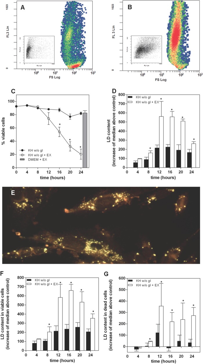
Under nutrient deprivation, inhibition of β-oxidation accelerates cell death and leads to the accumulation of LD. A and B, HeLa cells were kept for 10 h in KH without glucose and in the absence (A) or presence of 30 μm EX (B). After treatment, they were harvested and stained with PI and BODIPY 493/503 to measure viability and LD content. A and B show the event distribution for each treatment in terms of PI permeability (FL3) and size (FS). Inhibition of β-oxidation (B) induced death of nutrient-deprived cells. Insets show the event distribution of the dead cell population for each treatment in terms of FL3 and LD occurrence (FL1). The increased FL1 signal induced by EX is apparent. C, inhibition of β-oxidation with 30 μm EX accelerated the death of LN18 cells maintained in KH without glucose but had no effect on cells maintained in culture medium (DMEM), as denoted by a gray bar. Treatment with EX led to an overall accumulation of LD (D) that was very apparent in Nile red-stained cells after a 16-h treatment (E). Analysis of viable (F) and dead cells (G) shows increased LD occurrence in EX-treated samples. Results are means ± S.E. (error bars) of six independent experiments carried out with duplicate determinations. *, significantly different from KH without glucose.

