
FIGURE 2.
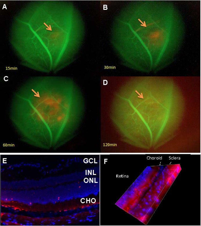
Periocular injected exosomes reach the choroid-retina. A–D, time course of the appearance of exosomes in the eye as seen by fluorescent microscopy. Mice preinjected with FITC-dextran, which labels blood vessels green, were injected periocularly with PKH26-labeled RAC-derived exosomes (10 μg) and examined in vivo using fluorescence microscopy at 15 (A), 30 (B), 60 (C), and 120 (D) min after injection (magnification, ×10). The arrows indicate PKH26-labeled (red) exosomes. E and F, confocal microscope of two-dimensional (E) and three-dimensional (F) view of frozen sections of the retina and choroid prepared after sacrifice of mice 60 min after periocular injection of RAC-derived exosomes labeled with PKH26 (red) (magnification, ×40; the blue stain of nucleus is DAPI. E, GCL, layer of ganglion cells; INL, inner nuclear layer; ONL, outer nuclear layer; CHO, choroid.