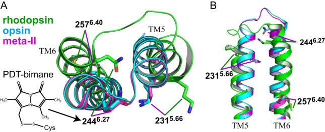
FIGURE 1.

Comparison of TM5 and TM6 in the crystal structures of opsin, rhodopsin, and photoactivated (meta-II) form. Cytoplasmic view (A) and side view (B) showing the relative displacement of TM5 and especially TM6 in the active (meta-II) and inactive forms of rhodopsin. Note that the crystal structure of opsin appears more like the active form of rhodopsin. Also indicated are the sites of the bimane labeling (position 2446.27), the quencher tryptophan (position 2315.66) used for the TrIQ studies discussed in the text, and a constitutively activating mutation M257Y (position 2576.40). The superscripts of the respective positions refer to Ballesteros-Weinstein numbering (18). The models were constructed using the structures of bovine opsin (light blue, PDB code 3CAP) (22), rhodopsin in the dark (green, PDB code 1GZM) (31), and rhodopsin in meta-II state (magenta, PDB code 3PQR) (10).
