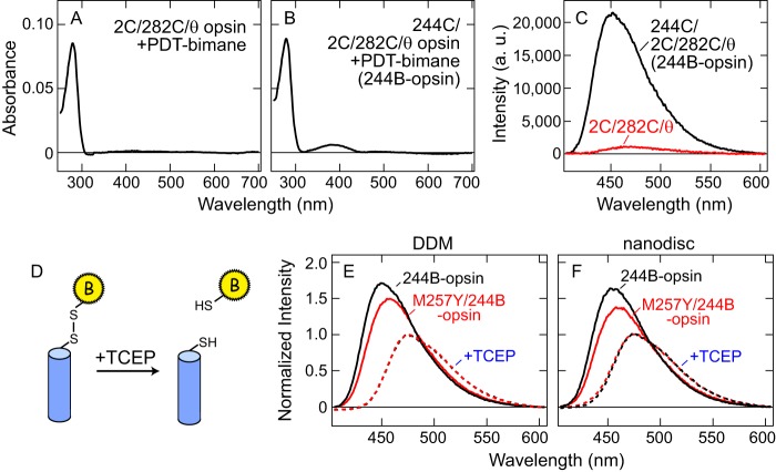
FIGURE 2.
PDT-bimane labeling on opsin mutants. A-C, example of specific incorporation of PDT-bimane label into position 244 in opsin mutant. Opsin mutants 244C/2C/282C/θ and 2C/282C/θ were labeled with PDT-bimane as described under “Experimental Procedures.” A, absorption spectrum of 2C/282C/θ opsin in DDM after labeling with PDT-bimane. B, absorption spectrum of 244C/2C/282C/θ opsin in DDM, after labeling with PDT-bimane (244B-opsin). C, fluorescence emission spectra using the same amount (500 nm) of both labeled opsins. Spectra clearly show that fluorescence of labeled 244C/2C/282C/θ opsin (244B-opsin, black) is much greater than that of labeled 2C/282C/θ opsin (red). This result indicates that PDT-bimane is specifically incorporated into Cys-244 not Cys-2/Cys-282 in opsin. Note that nonspecific bimane labeling on 2C/282C/θ opsin is less than 0.1 bimane/opsin. D, schematic representation of reduction of disulfide linkage between PDT-bimane and protein by a reducing reagent TCEP. E and F, normalization of fluorescence emission spectra by comparison to bimane emission after release from protein due to reduction of disulfide linkage. Solid lines indicate fluorescence emission spectra of 244B-opsin (black) and M257Y/244B-opsin (red) in DDM (E) and in nanodiscs (F). The red and black broken lines indicate the spectra of these same samples after release of the bimane label from the protein by reduction of the disulfide bond by TCEP (see “Experimental Procedures”). All fluorescence spectra are normalized to the fluorescence intensity after TCEP treatment.

