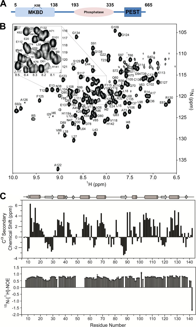
FIGURE 1.
The DUSP16 MKBD. A, domain architecture of DUSP16 including the N-terminal MKBD, the catalytic phosphatase domain, and the C-terminal PEST domain. B, fully annotated two-dimensional 1H,15N heteronuclear single quantum correlation spectrum of the DUSP16 MKBD. C, 13Cα chemical shift index of the DUSP16 MKBD; secondary structural elements of the crystal structure (PDB ID 2VSW) are shown (top panel). 15N,1H NOE of the DUSP16 MKBD, showing high rigidity of the structure throughout the protein (bottom panel).

