
FIGURE 1.
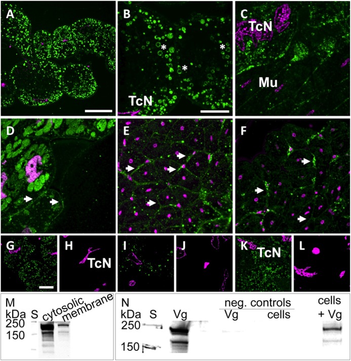
Vg binds to membranes and other cellular structures in the honey bee and to cells of an insect cell line. A–L, immunolabeling indicates the presence of Vg at vesicular (asterisks) and cellular membranes (arrowheads). Optical sections of abdominal (A–C) and head tissue (D–F) are labeled with anti-Vg (green) and DAPI nuclear stain (magenta). A–C, trophocytes are identified by irregularly shaped nuclei (TcN; see also G–L) and contain a large number of Vg-rich vesicles. Intense VgIR was also observed in other abdominal cell types (e.g. in muscle tissue (Mu)). D, hypopharyngeal glands with intense VgIR in Vg-rich granules and less intensely labeled cellular membranes (arrowheads). E and F, syncytial cells of muscle fibers with marked VgIR at their outer membranes (arrowheads). G–L, test sections treated with anti-Vg (G, I, and K) as compared with alternate negative control sections treated only with the secondary antibody (H, J, and L). The nuclear stain (DAPI) confirms that control and test images were taken from similar sites and cell types (trophocytes with characteristic TcNs). Scale bars, 50 μm (A), 20 μm (B–F), and 20 μm (G–L). M and N, Vg immunoblots detecting full-length Vg (180 kDa) and the 150-kDa Vg fragment present in the abdominal tissue (31). S, molecular weight standard. M, Vg and its 150-kDa fragment are found in both cytosolic and membrane fractions of abdominal honey bee tissue samples. N, Vg of abdominal protein extract binds to Sf9 insect cells during a 1-h incubation (last lane, cells + Vg). The first Vg lane is a positive control of abdominal protein extract. Negative controls were Vg incubated without cells (Vg) and cells without Vg (cells).