
FIGURE 4.
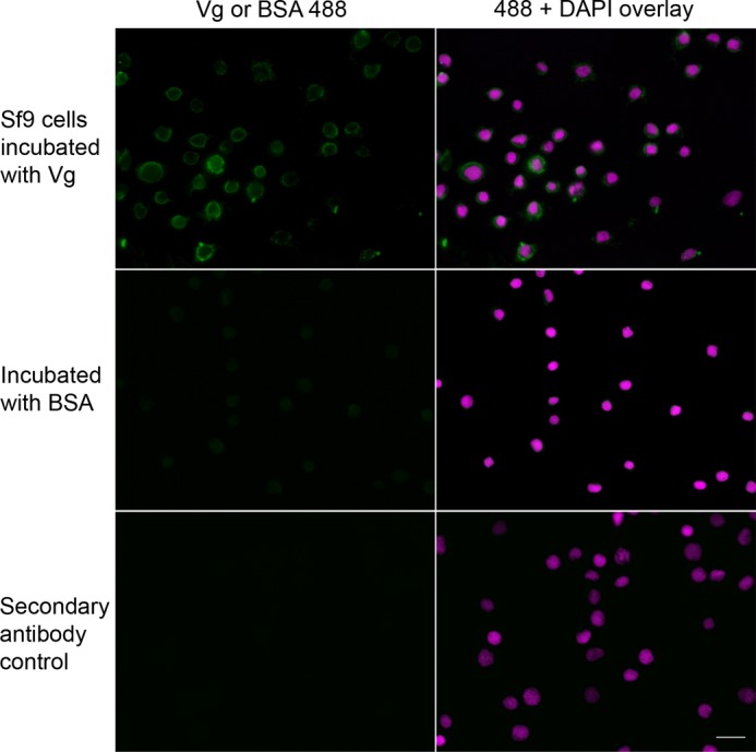
Vg binding to dead Sf9 cells compared with a BSA control. Killed Sf9 insect cells were incubated with Vg or BSA, followed by a treatment using rabbit anti-Vg or rabbit anti-BSA polyclonal primary antibody. The secondary antibody control was not incubated with a primary antibody, and, as expected, marked fluorescence was absent in these samples. All samples were treated with Alexa-488 (green) secondary antibody and DAPI. In contrast to Vg-incubated cells, the controls incubated with BSA did not exhibit marked positive staining, suggesting that binding of BSA was negligible. Scale bar, 20 μm.