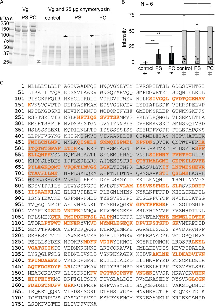
FIGURE 6.
A ∼120-kDa Vg segment is resistant to proteolysis in the presence of liposomes. A, a gel showing how a part of Vg (the band marked with black arrows) is not digested by chymotrypsin in the presence of PC or PS liposomes. The gray arrows show two bands, whose intensity in all lanes was used as a reference. S, molecular weight standard. Vg, untreated, purified Vg from honey bee abdomen. Control, no liposomes. PS, Vg with PS liposomes. PC, Vg with PC liposomes. B, the intensity of the bands marked with black arrows in A is significantly higher than the intensity of the corresponding gel region in the controls but does not differ between PS and PC. The reference bands (gray columns; gray arrows in A) showed no significant difference. The error bars show the S.D. of six band intensities. C, the LC-MS/MS hits (red) of the gel-extracted honey bee Vg segment that resists chymotrypsin digestion in the presence of liposomes (black arrows in A). The α-helical domain is highlighted gray. The most reliable peptide hits with high Mascot ion score (>75) are underlined.

