FIGURE 7.

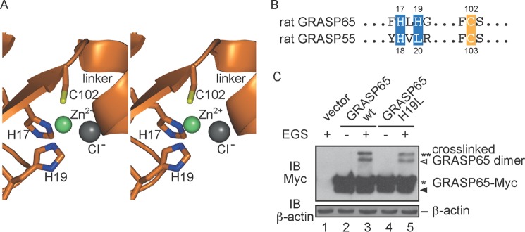
Zinc finger in GRASP65. A, stereoview of the zinc finger. Residues engaging the zinc ion are shown in stick representation. The zinc and chloride ions are shown as green and gray spheres, respectively. B, sequence alignment of the residues forming the zinc finger. C, HeLa cells were transfected with wild-type (wt) or mutant GRASP65-Myc and treated in the absence or presence of 400 μm EGS. The lysates were analyzed by SDS-PAGE and immunoblotting (IB) with anti-Myc antibodies. Asterisks indicate the number of GRASP65 molecules in the cross-linked product. The black arrowhead indicates degraded GRASP65, and the white arrowhead indicates cross-linked product containing degraded protein. The level of β-actin was monitored as a loading control.
