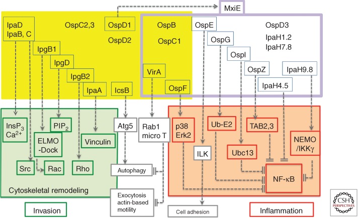
Figure 1.
The control of cell responses by Shigella T3S effectors. Shigella T3S effectors are shown in yellow and purple boxes. The T3S effectors boxed in blue represent those for which a function has been assigned. T3S effectors that are constitutively expressed (yellow box) or up-regulated following cell-contact induction of secretion (boxed in purple) and activation by MxiE. On cell contact, IpaB and IpaC insert into the host-cell membrane to form the “translocon,” required for the induction of InsP3-dependent Ca2+ responses. The targets of T3S effectors involved in Shigella invasion are boxed in green. IpaC, through its carboxy-terminal domain induces the recruitment and activation of the Src kinase. IpgB1 and IpgD, together with IpaC, participate in actin polymerization and membrane ruffle formation by targeting ELMO/Dock and hydrolyzing PIP2, respectively. IpgB2 that activates Rho, and IpaA that binds to vinculin, further reorganize the actin cytoskeleton to promote invasion. IcsB prevents autophagic responses by inhibiting binding of Atg5 to IcsA. The targets of T3S effectors that down-regulate inflammation are boxed in red. VirA prevents autophagy and IL-8 secretion through its GAP activity toward Rab1. VirA also inhibits microtubule polymerization to favor actin-based motility. Among the T3S effectors up-regulated by MxiE, OspF prevents the activation of the p38 and Erk2 MAP kinases via its phospho-threonine lyase activity. OspE reinforces cell adhesion by targeting ILK. The targets of OspG, OspI, OspZ, IpaH4.5, and IpaH9.8 that down-regulate NF-κB activation are indicated.

