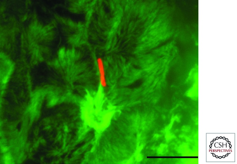
Figure 2.
Filopodial capture and Shigella invasion at the apical side of polarized Caco-2 cells. Shown is a Z projection of confocal fluorescence images of Shigella-induced actin foci at the apical side of polarized Caco-2 cells. Red, anti-LPS immunostaining; green, Alexa488-phalloidin staining. Z sections were acquired via spinning-disk confocal microscopy. Scale bar, 5 mm.

