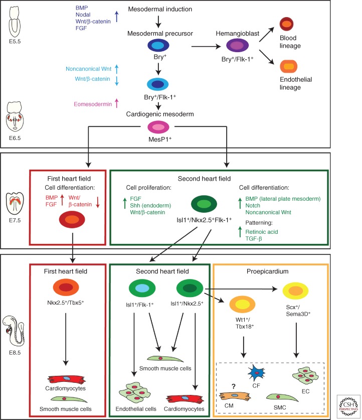
Figure 2.
Cellular hierarchy of cardiac progenitor cells and their lineage specification. Several signaling pathways (BMP, Nodal, Wnt/β-catenin, FGF) interact to induce mesoderm; Brachyury (Bry) positive mesodermal precursors first differentiate through Bry+/Flk-1+ hemangioblasts toward endothelial and blood-cell lineages (around E5.5 during mouse development). Slightly later during development after down-regulation of Wnt/β-catenin signaling and induction of noncannonical Wnt signals a second wave of Bry+/Flk-1+ mesodermal progenitors appears. Eomesodermin signaling drives cardiogenic mesoderm specification from these primitive mesodermal precursors. Cardiogenic mesoderm is marked by the expression of mesoderm posterior 1 (Mesp1) (around E6.5 in mouse embryogenesis). Early mesoderm-derived cardiac precursors undergo further lineage restriction and differentiate into progenitor pools that populate the FHF and SHF, respectively. At this stage (E7.5 mouse development) FHF progenitors start to differentiate upon BMP and FGF action toward cardiomyocytes and smooth muscle cells, whereas Wnt/β-catenin, FGF, and endodermal Shh signaling keeps SHF progenitors in a proliferative state. These SHF progenitors are defined by the molecular signature Isl-1+/Nkx2.5+/Flk-1+. SHF progenitors are now gradually added to the looping heart tube and get further restricted in their differentiation potential (E8.5). Two subpopulations of SHF progenitors can be distinguished. One population marked by the expression of Isl-1 and Flk-1 differentiates into endothelial cells and smooth muscle cells, whereas a second pool of Isl-1+/Nkx2.5+ SHF precursors provides smooth muscle cells and cardiomyocytes as well as contributing to the proepicardial lineages (Wt1+/Tbx18+ and Scx+/Sema3D+ populations), which later form cardiac fibroblasts (CF), smooth muscle cells (SMCs), endothelial cells (EC), and cardiomyocytes (CM), with the latter contribution being still unclear. These distinct SHF progenitor populations differentiate upon BMP signals from the lateral plate mesoderm as well as Notch and noncanonical Wnt signals. SHF patterning is governed by RA and TGF-β signals.

