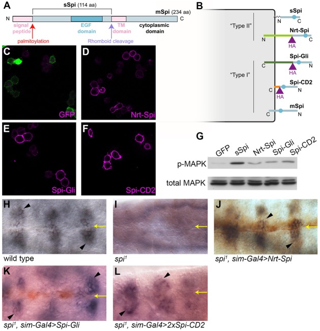
Fig. 1.
Transmembrane Spi chimeras are trafficked to the cell surface and can activate the EGFR in vitro. (A) Full-length mSpi pro-protein has its receptor-binding EGF domain located close to the transmembrane (TM) domain. Removal of the signal peptide and cleavage in the transmembrane domain by Rho generates active sSpi, which is palmitoylated at its N-terminus. (B) Chimeric transmembrane proteins were created by fusing sSpi to Nrt (light green), Gli (dark green) or the transmembrane domain of CD2 (orange). All three chimeras have an HA tag (purple). The EGF domain is represented by a blue circle. (C–F) S2R+ cells expressing Nrt–Spi (D), Spi–Gli (E), Spi–CD2 (F) or control cells expressing only intracellular GFP (C) were stained for HA (magenta, D–F) or GFP (magenta, C) in the absence of detergent. GFP fluorescence (green) is visible in C. (G) S2 cells expressing intracellular GFP, sSpi, Nrt–Spi, Spi–Gli or Spi-CD2 were co-cultured with EGFR-expressing D2F cells and assayed by western blotting for MAPK phosphorylation (p-MAPK) and total MAPK levels. (H–L) FasIII expression (purple, arrowheads) in the embryonic ventral ectoderm (H) was lost in spi zygotic mutant embryos (I) and was rescued by expression of Nrt–Spi (J), Spi–Gli (K) or two copies of Spi–CD2 (L)(stained with anti-HA antibody in brown) driven in the ventral midline cells by sim-GAL4. Yellow arrows indicate the ventral midline.

