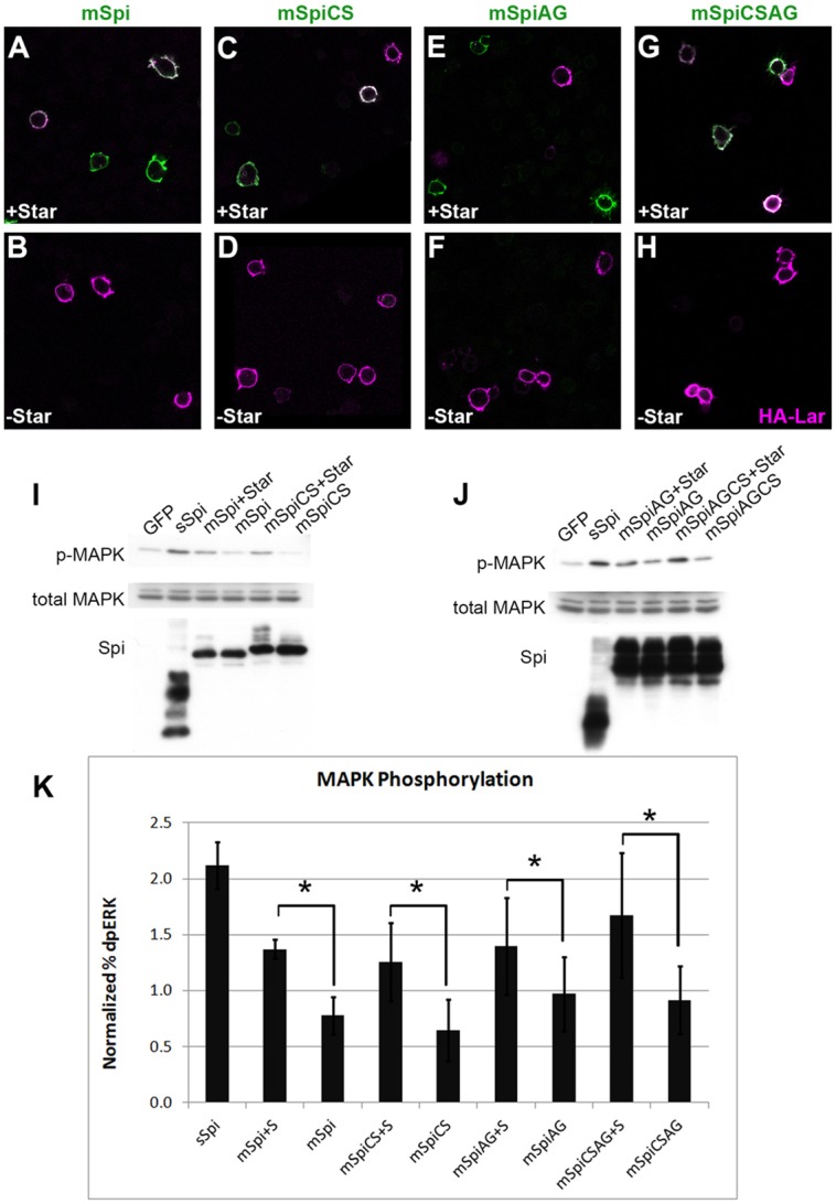
Fig. 4.

mSpi pro-protein activates the EGFR in vitro. (A–H) Flag-tagged mSpi (A,B), mSpiCS (C,D), mSpiAG (E,F) or mSpiCSAG (G,H) was detected on the surface of S2 cells by anti-Flag staining (green) in the absence of detergent when co-expressed with Star (A,C,E,G). In the absence of Star, no cell-surface mSpi was detected (B,D,F,H). Staining for the transmembrane protein HA–Lar (magenta) marks the plasma membrane. (I–K) Co-culture of S2 cells expressing mSpi, mSpiCS, mSpiAG or mSpiCSAG with EGFR-expressing D2F cells resulted in MAPK phosphorylation. Quantification was performed using Li-Cor Odyssey (K). For each condition, the phospho-MAPK level was divided by total MAPK level (% dpERK) and this value was normalized to the percentage dpERK obtained for control D2F cells co-cultured with S2 cells expressing intracellular GFP alone in that experiment. The mean±s.e.m. of three experiments is shown. sSpi-expressing cells induced more than a twofold increase in phospho-MAPK in co-cultured D2F cells compared to GFP-expressing cells. Each mSpi construct co-expressed with Star induced about a 1.5-fold increase in phospho-MAPK compared to GFP, whereas mSpi constructs without co-expressed Star did not increase phospho-MAPK above the level observed with GFP. *P<0.05 between the percentage dpERK induced by each Spi construct when co-expressed with Star compared to the percentage dpERK induced when expressed without Star (asterisks) as determined by paired Student's t-tests.
