Fig. 4.

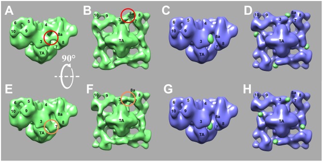
Three-dimensional reconstruction of RyR2R3595-GFP and RyR2K4269-GFP. (A,B,E,F) Surface representations of the 3D reconstruction of RyR2R3595-GFP (A,B) and RyR2K4269-GFP (E,F), shown in a side view (A,E) and in bottom view (B,F). The side view shows the surface that would be perpendicular to the plane of the sarcoplasmic reticulum membrane, and the bottom view shows the surface parallel to the sarcoplasmic reticulum membrane, which in situ would face the sarcoplasmic reticulum lumen. Red and orange circles highlight the locations of the extra masses representing GFP. (C,D,G,H) Surface representations of the 3D reconstruction of RyR2 control shown in blue, and the difference map [subtraction of 3D volume of RyR2 control from that of RyR2R3595-GFP (C and D) or RyR2K4269-GFP (G and H)] shown in green and superimposed on the 3D reconstruction of RyR2 control.
