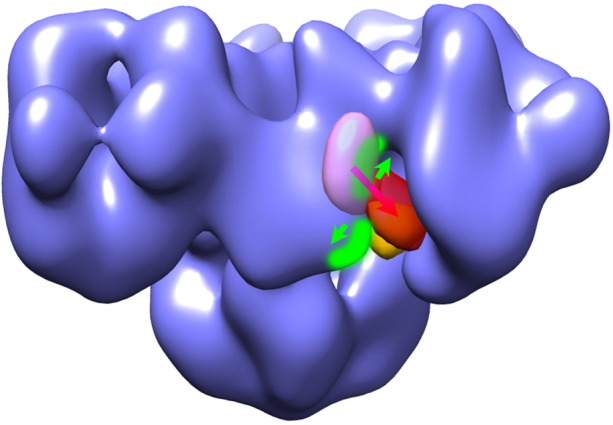
Fig. 6.

Hypothetical model of RyR conformation changes in the CaM-binding region and translocation of CaM when the RyR channel switches from closed to open. The transparent purple mass represents the apo-CaM-binding site, and the transparent red mass is the Ca2+-CaM-binding site in RyR1. The orange mass indicates the apo-CaM-binding site in RyR2. Two structural domains bearing CaM-binding sequences 3615–3644 and 4303–4328 in RyR1 are highlighted in green. Green arrows demonstrate that the two domains move apart when the RyR channel is activated, and the purple-red arrow indicates the shift of CaM from the apo-binding site to the Ca2+ binding site that occurs in RyR1 but not in RyR2. Figure adapted from Samsó and Wagenknecht and Huang et al., with modifications (Samsó and Wagenknecht, 2002; Huang et al., 2012).
