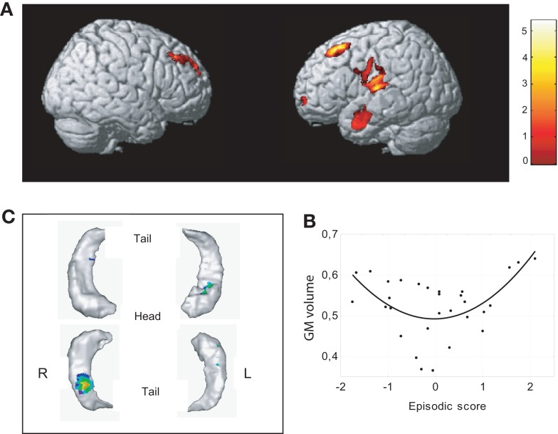
Figure 4.
Neural correlates of associative memory. (A) Significant positive correlation (quadratic, U-shaped) with episodic score was found with the volume of gray matter in dorsolateral frontal regions bilaterally, the superior temporal cortex, the anterior middle temporal gyrus, and the ventrolateral prefrontal cortex (upper figure, uncorrected p < 0.001, K > 500). (B) Scatterplots of episodic effects are shown for regions identified in whole brain analyses. (C) Significant positive correlation (quadratic, U-shaped) was observed between memory and right hippocampal body and the anterior part of the left hippocampus (uncorrected p < 0.01, K > 100).

