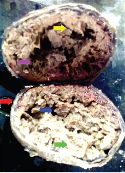
Figure 2.

On cut section, the tumor revealed a thickened capsule of size 1.2 cm [red arrow]. The entire tumor mass underneath the capsule showed extensive necrosis, cystic degeneration [green arrow] and hemorrhagic areas forming dry layers, like those of thin nets [pink] and septae [yellow], containing empty spaces [blue arrow] within them
