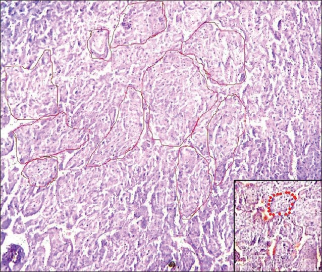
Figure 3.

H and E section under light microscopy (×10) shows large, oval cells arranged in organoid and trabecular pattern separated by a delicate vascular network, which appear as red dots in a magnified view (see inset). The whole appearance, which is classically described as Zellballen pattern, meaning ‘well-defined nests,’ is prominent with encircling red lines
