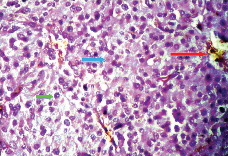
Figure 4.

H and E section under light microscopy (×40) shows tumor cells have abundant acidophilic cytoplasm containing granules [blue arrow] with moderate variation in nuclear size and shape, some showing inclusion like structures [green arrow] and insignificant mitosis. Areas of necrosis, hemorrhage [red arrow] and cystic degeneration
