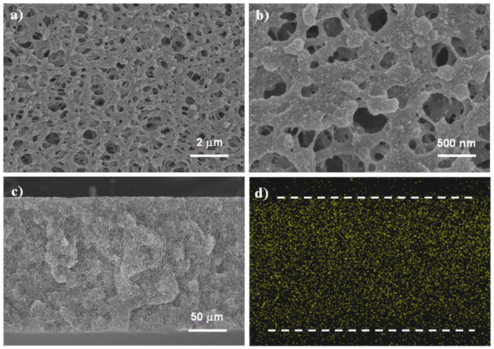
Figure 1. Morphology of the mineral-coated membranes prepared by grafting PAA and then depositing CaCO3 nanoparticles on polypropylene microfiltration membranes.
(a) SEM image of the membrane surface in large-area view. (b) Enlarged view of the membrane surface. (c) SEM image of the membrane cross-section. (d) Distribution of calcium element (points measured by EDX mapping analysis) on the membrane cross-section (The dash lines indicate the edges of the cross-section. The dense spots below the lower dash line originate from the bottom surface of the membrane).

