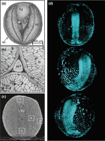
Fig 1.

Structural organization of a developing oilseed rape (Brassica napus) seed using conventional and non-invasive analysis. (a) Horizontal mid section of a developing seed sampled c. 30 d after flowering. (b) A TEM acquired image shows the presence of lipid bodies inside the cotyledonary cells and extracellular voids at the cell corners. (c) A high resolution CT vertical radial section of a whole rapeseed (1.7 μm per pixel). A fuller visualization of these images is provided in Supporting Information Movie S1. The rectangles indicate regions modelled in Figs 2 and 3. (d) A rotation series of views of the void spaces in a seed obtained from high resolution CT. An animated three-dimensional model is given as Movie S2. as, airspace; cw, cell wall; el, endospermal liquid; hy, hypocotyl; lb, lipid body; ic, inner cotyledon; oc, outer cotyledon; sc, seed coat (testa).
