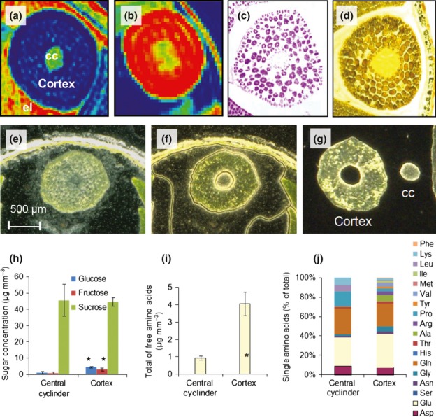
Fig 7.

Histological and biochemical analysis of the oilseed rape (Brassica napus) hypocotyl. (a) Water and (b) lipid distribution as determined by MRI (low levels marked by blue, high levels by red). (c) Cruciferin distribution, as determined by immunostaining. (d) Starch distribution, as determined by iodine staining. (e–g) For laser micro-dissection procedure. (e) An intact section of the hypocotyl. (f) Tissue regions dissected by laser. (g) Dissected stele and cortex region of the hypocotyl. (h–j) Laser-dissected material used for the analysis of sugars (h) and free amino acids (i, j). *, Statistically significant differences between the cortex and the stele (t-test, P < 0.05, n = 6). cc, central cylinder; el, endospermal liquid.
