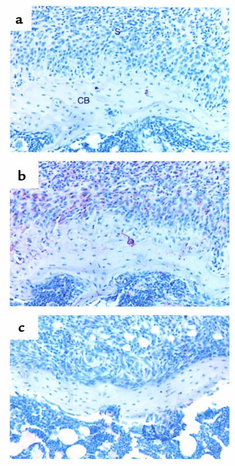
Figure 11.
Effects of IL-4 on OPGL expression in the synovium. (a and b) Arthritic knee joint of a mouse 7 days after intra-articular injection of 1.107 pfu of Ad5del70-3 control vector showing staining for OPGL, detected with a control anti-goat Ab (a) or the anti-RANKL Ab (b). Note the OPGL expression in the synovium and in cells along the cortical bone. (c) Knee joint of a mouse 7 days after intra-articular injection of 1.107 pfu of Ad5E1mIL-4 showing staining for OPGL, detected with the anti-RANKL Ab. Note the decreased OPGL expression in the synovium and in cells along the cortical bone. a–c, ×250. S, synovium; CB, cortical bone.

