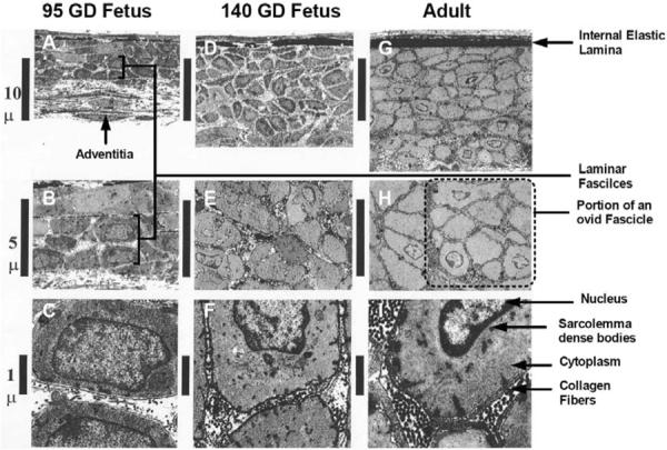
Fig. (1).

Transmission electron microscopy images of arterial walls of main branch middle cerebral arteries of preterm 95-gestational day fetus, near-term 140-gestational day fetus, and adult sheep. Original magnification of axial longitudinal section: ×1,600 (A, D, and G), ×8,000 (B, E, and H), and ×10,000 (C, F, and I) [10].
