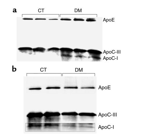
Figure 2.
(a) Western blot analysis of isolated VLDL from CT and DM C57BL/6 mice for apoE, apoC-III, and apoC-I. Constant amounts of protein were loaded. ApoE, apoC-III, and apoC-I are increased in DM, apoC-I most prominently. (b) Composite of blot of isolated HDL separately developed for these antibodies. In contrast, apoE, apoC-III, and apoC-I are modestly decreased in DM.

