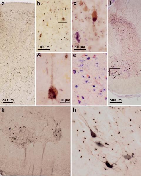
Figure 1. pTDP-43 pathology in the motocortex and spinal cord.
a. ALS-associated pTDP-43 pathology was observed in Betz pyramidal cells of layer Vb and in smaller projection neurons of layers II, III, V, and VI in the primary motor area (Brodmann area 4) (case 75, stage 4). b. Layer Vb of the primary motor cortex with affection of Betz pyramidal cells (case 75, stage 4). c. Detail micrograph (framed area in b) shows single Betz cell in higher resolution with pTDP-43 aggregates detectable in the somatodendritic compartment and in unmyelinated initial portions of the axon. d. Pigment-Nissl stain of layer Vb of the motor cortex depicting a Betz pyramidal cell (upper right) and loss of a Betz cell indicated by lipofuscin pigment remnants in the neuropil (pigment incontinence, lower left) (case 14, stage 2). e. Pigment-Nissl stain of spinal cord ventral horn (framed area in f is rotated 90° to the right in e) depicting α-motoneurons alongside neuronal loss indicated by lipofuscin pigment remnants in the neuropil (case 13, stage 2). f. Pigment-Nissl overview of spinal cord ventral and dorsal horns. g,h. Spinal cord α-motoneurons of layer 9 showing pTDP-43 aggregates in neuronal cell bodies as well as in dendrites and axons (heading towards the ventral root) (case 48, stage 3). Note affected oligodendrocytes in h (case 38, stage 3). Scale in a applies to g; scale bar in b is also valid for e; scale bar in d applies to h.

