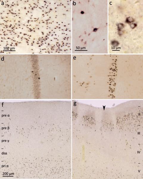
Figure 3. pTDP-43 pathology in the striatum, anteromedial fields of the temporal lobe and the hippocampal formation.
a. Severe pTDP-43 pathology in striatum involving mainly medium-sized projection neurons (case 69, stage 4). b. Micrograph of a group of medium-sized projection neurons and two large cholinergic local circuit neurons of the striatum showing ALS-associated lesions (case 70, stage 4). c. Immunoreactive medium-sized spiny projection cells of the striatum at higher magnification (case 70, stage 4). d. Mild involvement of hippocampal formation with pTDP-43 pathology in few granular cells of the dentate fascia (case 60, stage 4). e. Marked pTDP-43 pathology in granular cells of the dentate fascia (case 69, stage 4). Uninvolved plexiform layer and portion of the fourth sector of the Ammon’s horn is seen in the right halves of d and e. f. pTDP-43 pathology in pyramidal cells of the entorhinal region, mainly affecting layers pre-β and pri-α. Borders of allocortical entorhinal layers are indicated at the left (diss – lamina dissecans, pre – layers of the external main stratum, pri – layers of the internal main stratum) (case 69, stage 4). g. Transition between entorhinal region (left) and transentorhinal region (right) is indicated by large arrowhead. pTDP-43 pathology in pyramidal cells of the transentorhinal region affects neocortical layers: here, borders of neocortical layers II-V (at right) (case 69, stage 4). Scale bar in a also applies to d and e; scale bar in f is also valid for g. Paraffin-embedded 70 μm sections.

