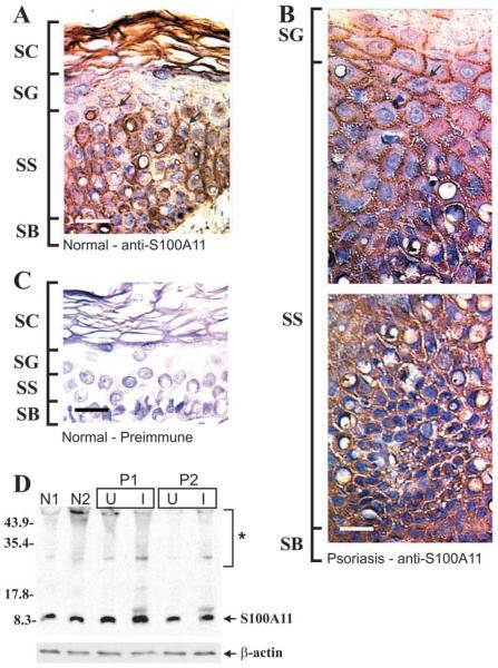
Figure 7.
S100A11 in epidermis. Epidermal sections were prepared and stained with rabbit anti-human S100A11. (A) Normal epidermis stained with anti-S100A11. (B) Involved psoriatic epidermis stained with anti-S100A11. (C) Normal epidermis stained with preimmune serum. (D) Equivalent amounts of whole-cell extract (based on protein) prepared from normal epidermis (patients N1, N2), and psoriatic epidermis (patients P1, P2) were electrophoresed on a 12% acrylamide gel. Samples from psoriasis patients include uninvolved (U) and involved (I) tissue. Primary antibody binding was visualized using an appropriate secondary antibody and chemiluminescence detection agents. β-Actin was detected as a control to confirm equal loading. Asterisk indicates high molecular weight anti-S100A11 immunoreactive bands (Ruse et al. 2001). Arrows (A,B) indicate membrane-associated S100A11. Bar = 10 μm.

