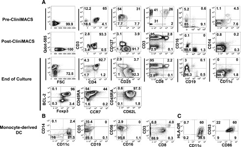
Figure 3.

Phenotypic characterization of Tregs and monocyte-derived DCs from leukapheresis. (A) Phenotype of Tregs from donor leukapheresis before and after enrichment and after large-scale expansion. Mononuclear cells were obtained from blood cell apheresis followed by positive selection with CD25 paramagnetic beads for immunoabsorption on column by CliniMACS. The top row (pre-CliniMACS) shows phenotype of mononuclear cells after donor apheresis. The second row (post-CliniMACS) shows viability, phenotype, and purity of Tregs after CliniMACS separation. The third and fourth data rows show Treg viability, phenotype, and purity after 12-day ex vivo expansion using DCs from an HLA-identical sibling, IL-2, IL-15, and rapamycin. Viability was assessed by Qdot 585-A exclusion, and viable cells were gated for marker studies. CD3+/CD4+ double-positive cells were gated for assessment of CD25 and CD127 expression. CD25+/CD127− cells were gated for assessment of Foxp3, BCL-2, CD45RA, and CCR7 expression. Cells other than CD4 from the end-of-culture population were also tested. Cell separation and culture was performed in the cell therapy core facility at Moffitt Cancer Center. (B,C) Surface phenotype of cells enriched for monocyte-derived DCs before coculture with Tregs. All flow-cytometric plots are gated on live cells, and HLA-DR, CD86, and CD83 plots are also gated on CD11c+/CD14− cells.
