
Figure 2.
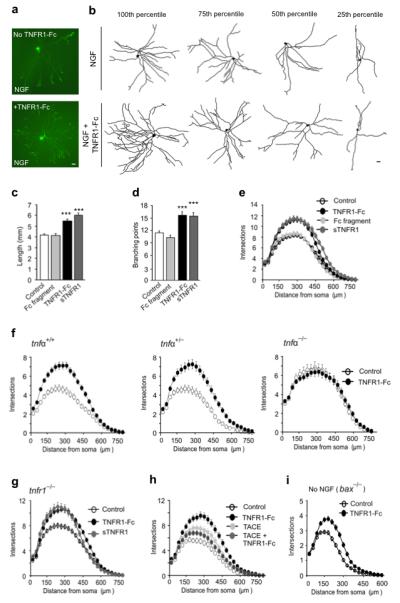
TNFR1-promoted TNFα reverse signaling enhances neurite growth from SCG P0 neurons. (a) Representative neurons cultured for 24 h in medium containing 10 ng/ml NGF, with or without 10 ng/ml TNFR1-Fc. (b) Representative camera lucida drawings of neurons corresponding to the 100th, 75th, 50th and 25th percentiles in terms of total neurite length. Scale bars = 20 μm. Length (c), branch point number (d) and Sholl profiles (e) of neurons cultured for 24 h with either NGF alone (control, n = 342) or NGF plus 10 ng/ml of human Fc fragment (n = 228), 10 ng/ml TNFR1-Fc (n = 171) or 5 μg/ml sTNFR1 (n = 180). (f) Sholl plots of neurons from P0 tnfα+/+, tnfα+/− and tnfα−/− littermates cultured for 24 h with NGF alone (control) or NGF plus TNFR1-Fc (n = 150). (g) Sholl plots of neurons from tnfr1−/− mice grown for 24 h with either NGF alone (control) or NGF plus TNFR1-Fc or sTNFR1 (n = 150). (h) Sholl plots of neurons cultured for 24 h with either NGF alone (control, n = 234) or NGF plus TNFR1-Fc (n = 243), 200 ng/ml TACE (n = 247) and TNFR1-Fc plus TACE (n = 248). (i) Sholl plots of the neurite arbors of neurons from bax−/− mice grown for 24 h in the presence (n = 174) or absence (n= 196) of TNFR1-Fc in medium lacking NGF. Mean ± s.e.m of data from 3 to 5 separate experiments of each type (*** indicates P <0.001, statistical comparison with control).