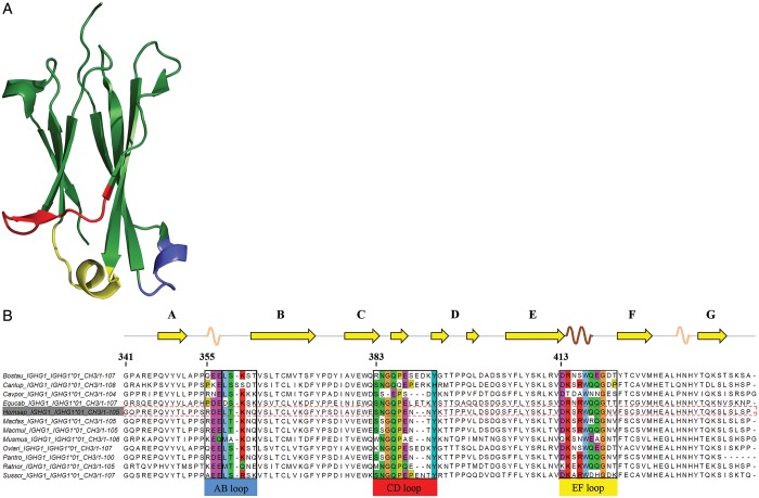
Fig. 1.
(A) Crystal structure of the CH3 domain of human IgG1 (PDB code 1OQO), showing the typical immunoglobulin fold composed of two β-sheets formed by three and four β-strands, respectively. The loops connecting these β-strands at the C-terminus of the CH3 domain are shown in blue (AB-loop), red (CD-loop) and yellow (EF-loop). (B) Multiple sequence alignment of IgG1-CH3 of 12 species. Loop regions are colored according to the ClustalW color code in order to emphasize possible conservation of amino acid side chain character (Larkin et al., 2007).

