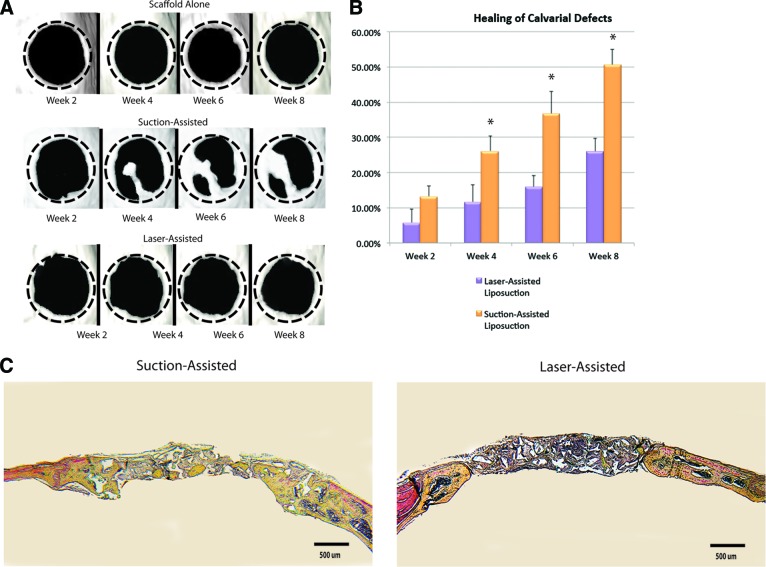
Figure 5.
Application of adipose-derived stromal cells in calvarial defects. (A): Three-dimensional reconstruction of calvarial defects. Mice were scanned at 2, 4, 6, and 8 weeks following surgery. (B): Quantification of osseous healing by micro-computed tomography revealed significantly more healing with adipose-derived stromal cells isolated from adipose tissue harvested via suction-assisted liposuction relative to laser-assisted liposuction (*, p < .05) at the 4-, 6-, and 8-week time points. (C): Calvarial defects 4 mm in size were allowed to heal for 8 weeks before histological analysis by Movat's pentachrome staining. Pictures were taken in the middle of the defect site. In pentachrome stains, bone appears yellow.

