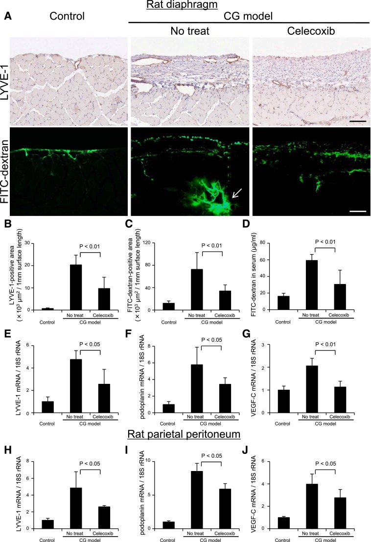
Figure 11.
Drainage of FITC dextran administered into the abdominal cavity is suppressed by inhibition of lymphangiogenesis with cyclooxygenase-2 inhibitor. (A, upper panels) Immunohistochemical staining of LYVE-1 in the diaphragm of control rats, celecoxib-treated (daily oral administration of 50 mg/kg body wt) CG rats, and untreated CG rats. (A, lower panels) Rats were given intraperitoneal injections of 50 mg FITC dextran (molecular weight=2,000,000). Twenty minutes later, blood, peritoneal, and diaphragmatic samples were obtained. Immunohistochemical findings in the diaphragm of control, untreated, and celecoxib-treated rats were recorded. Scale bars, 100 μm. Arrow indicates the accumulation of FITC dextran in the central collector of lymphatic vessel. Quantification of positive area determined by MetaMorph 6.3 image software (Universal Imaging, West Chester, PA) in the diaphragm for (B) LYVE-1 and (C) FITC dextran showed that celecoxib treatment significantly reduced both positive areas compared with the untreated CG rats. (D) To assess the amount of FITC dextran in blood drawn at euthanization, absorbance of serum samples was measured at 493 nm by spectrophotometry. For preparation of the standard curve, FITC dextran was reconstituted and diluted with normal rat serum. Concentrations of FITC dextran in the serum were significantly decreased in celecoxib-treated CG rats compared with untreated CG rats. Real-time PCR analysis of (E and H) LYVE-1, (F and I) podoplanin, and (G and J) VEGF-C mRNA in (E–G) the rat diaphragm and (H–J) the parietal peritoneum indicated that increased expression of LYVE-1, podoplanin, and VEGF-C mRNA in CG rats was significantly suppressed by celecoxib treatment. Control, n=5; CG models with no treat, n=6; CG models treated with celecoxib, n=6.

