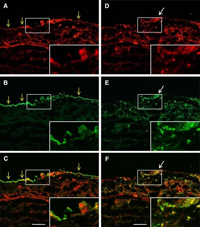
Figure 3.
Double immunofluorescent analysis shows VEGF-C expression by mesothelial cells and macrophages in human peritoneal biopsy specimens of bacterial peritonitis. Biopsy specimens were (A and D) stained with VEGF-C and costained with (B) the mesothelial cell marker cytokeratin or (E) the macrophage marker CD68. Respective merged images are shown in C and F. Cytokeratin-positive mesothelial cells (yellow arrows) and CD68-positive macrophages (white arrows) both expressed VEGF-C. (Insets) Magnification of the white boxed area. Scale bars, 100 μm.

