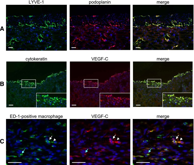
Figure 7.
Double immunofluorescent staining of frozen sections of a CG rat diaphragm shows that VEGF-C is expressed by cytokeratin-positive mesothelial cells and ED-1–positive macrophages. (A) The expression pattern of LYVE-1 (green) was similar to the expression pattern of podoplanin (red). (B) VEGF-C (red) was expressed in cytokeratin-positive mesothelial cells (green). (C) VEGF-C (red) was expressed in ED-1–positive macrophages (green). Arrows and arrowheads of the same color indicate the same cells. Nuclei were counterstained with 4',6-diamino-2-phenylindole (blue). (Insets) Magnification of the white boxed area. Scale bars, 50 μm.

