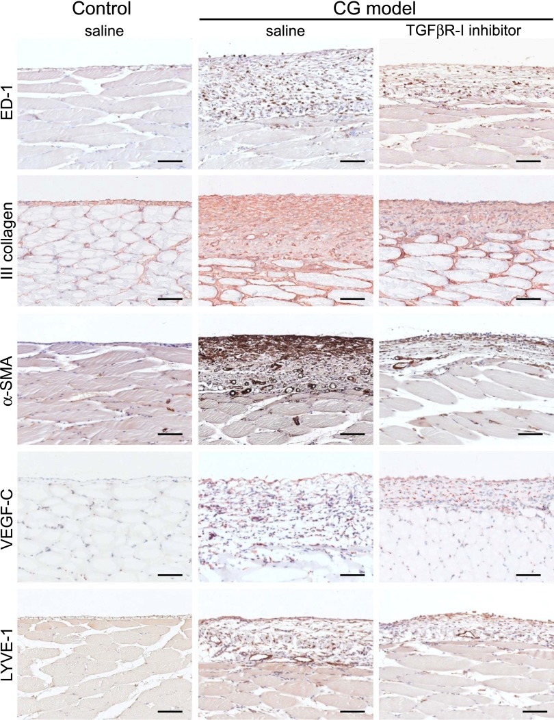
Figure 8.
Under immunohistochemical analysis of the parietal peritoneum, TGFβR-I inhibitor suppresses fibrosis and lymphangiogenesis. Histochemical staining of ED-1–positive macrophages, type III collagen, α-SMA, VEGF-C, and LYVE-1 in TGFβR-I inhibitor-treated (daily intraperitoneal injection of 3 μg/g body wt) and untreated CG model rats and control rats. Scale bars, 100 μm.

