Figure 6.
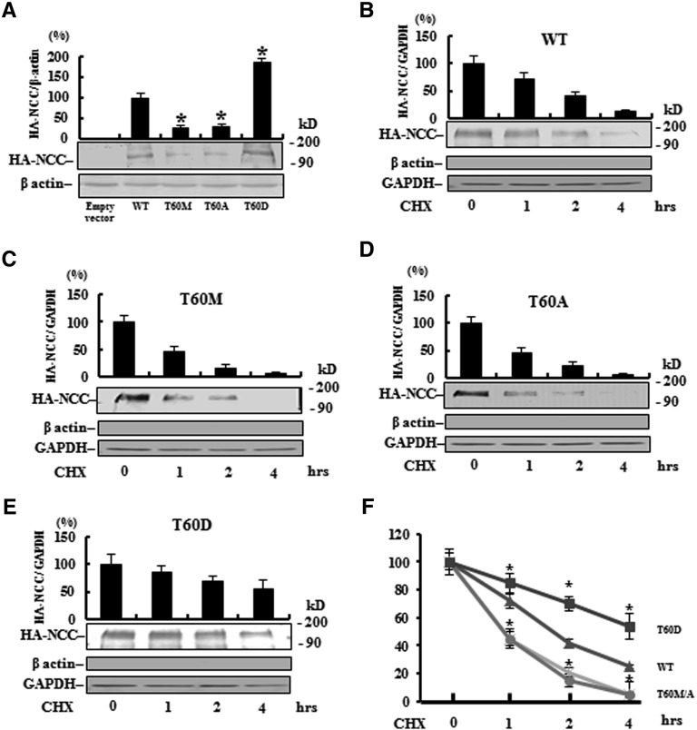
Phosphorylation on T60 affects total NCC expression and membrane stability in MDCK cells. (A) Semiquantitative (n=3) (upper panel) and a representative immunoblot (lower panel) of total NCC protein abundance and (B–E) change in membrane abundance of WT, T60M, T60A, and T60D NCC, respectively, after cycloheximide (CHX) treatment in MDCK cells. The lack of β-actin but positive glyceraldehyde 3-phosphate dehydrogenase (GAPDH) suggests that the loaded protein is obtained from the membrane fraction. (F) Compared with WT, T60A/M NCC exhibits more rapid degradation but T60D NCC has greater membrane stability. *P<0.05 versus WT.

