Figure 1.
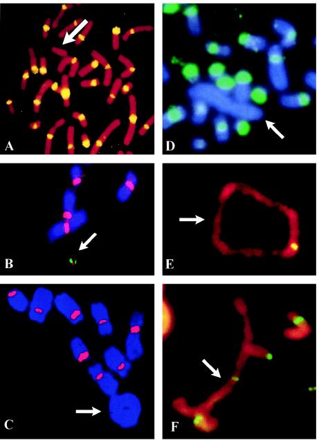
FISH analysis of human neocentric chromosomes. A, Patient cells with a 10q25 neocentromere-containing mardel(10) chromosome (arrow), using pancentromeric α-satellite probe, demonstrating absence of α-satellite (yellow) on the marker chromosome. Image taken from Voullaire et al. (1993). B, A stable, <2-Mb HAC (arrow) engineered from the mardel(10) chromosome shown in A (Saffery et al. 2001). Chromosome staining is with DAPI. Image courtesy of L. Wong. C–F, Partial metaphases of well-differentiated liposarcoma cases, using FISH with a pancentromeric α-satellite probe (C and D) and immunostaining with anticentromere antibody (E and F). Arrows indicate the supernumerary rings and large rod marker chromosomes. FISH signals (red in C or green in D) with the α-satellite probe are observed on all chromosomes except the supernumerary ring (C) and large marker (D). Positive staining with the anticentromere antibody (yellow or green) is observed on all chromosomes including the supernumerary analphoid ring (E) and large marker (F). Images courtesy of F. Pedeutour and N. Sirvent.

