Figure 7.

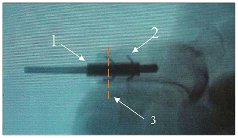
Animal 2. X-ray of pylon “1” (see Figure 5(c)) with side elements “2” after implantation to bone with the precut slots (see Figure 6). Red dashed line “3” shows plane of histology cross section (see Figure 10).

Animal 2. X-ray of pylon “1” (see Figure 5(c)) with side elements “2” after implantation to bone with the precut slots (see Figure 6). Red dashed line “3” shows plane of histology cross section (see Figure 10).