
Fig. 1.
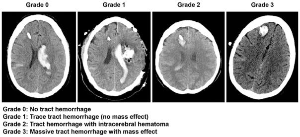
EVD tract hemorrhage grading scale (grading scale was defined for the purpose of this study). Examples of right frontal EVD’s (noncontrast head CT’s). The Grade 0 example is a patient with no tract hemorrhage, who had intraventricular hemorrhage primarily in left lateral ventricle and some right subarachnoid hemorrhage. The Grade 1 example is a patient with a intraparenchymal hematoma (left subcortical) status post decompressive hemicraniectomy and hematoma evacuation, large left lateral ventricular clot and trace cortical subarachnoid hemorrhage. There is trace hemorrhage around the EVD tract. The Grade 2 example is a patient with hematoma formation without midline shift (mass effect) with intraventricular blood and subarachnoid hemorrhage. The Grade 3 example shows sizeable tract hematoma with surrounding hypodense cerebral edema with subtle midline shift of the right parasagittal brain from right to left