
Fig. 3.
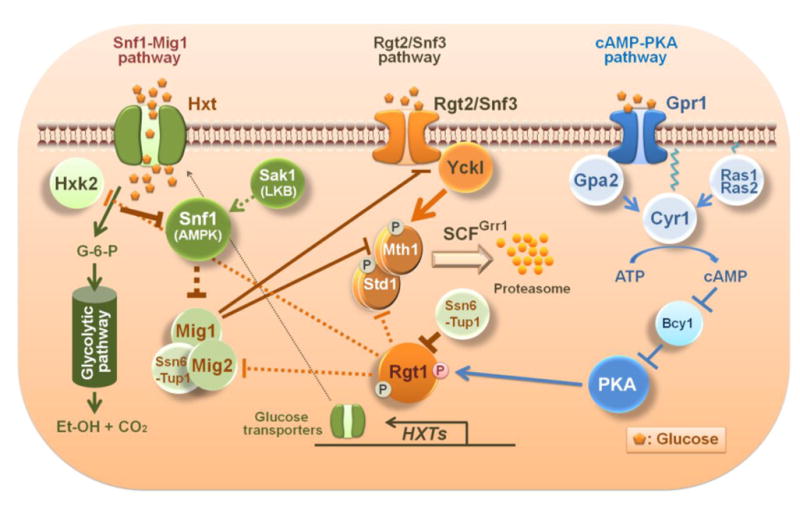
Schematic diagram of the crosstalk between glucose signaling pathways in yeast. Yck I (Yck1 and Yck2) phosphorylates Mth1 and Std1 upon activation by glucose-bound Rgt2 and Snf3 glucose sensors. Phosphorylated Mth1 and Std1 are ubiquitinated by the SCFGrr1 complex and degraded by the proteasome. The PKA phosphorylation sites in the amino terminal region of Rgt1 are exposed and available for phosphorylation when Mth1 is degraded. Phosphorylated Rgt1 is dissociated form Ssn6-Tup1 and subsequently from DNA, leading to derepression of Rgt1 target genes, such as the HXT and HXK2 genes. The Rgt2/Snf3 pathway regulates itself through glucose-induction of STD1 gene expression. Consequently, the STD1 gene is expressed at the same time that the Std1 protein is degraded in response to glucose [13]. By contrast, glucose stimulates Mth1 degradation but also represses Mth1 expression via Mig1 and Mig2. Glucose uptake is required for the generation of the glucose repression signal that leads to inactivation of the Snf1 kinase [18]. Expression of the MIG2 gene is induced by glucose via the Rgt2/Snf3 pathway. Glucose-repression of SNF3 gene expression by Mig1 reflects the probable function of Snf3 as a high affinity glucose sensor, representing another important feature of the interaction between the glucose induction and repression pathways.