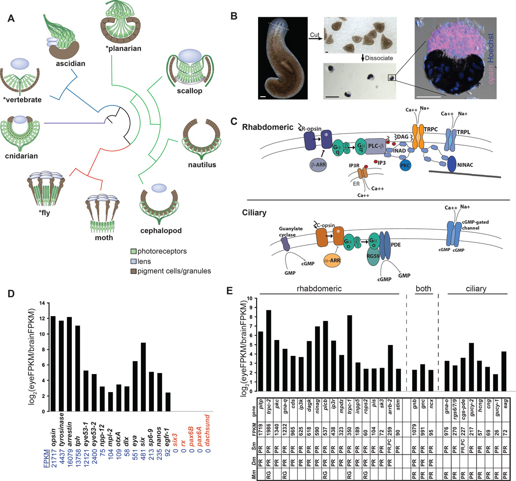
Figure 1. cDNA sequencing of purified planarian eyes.
(A) Schematic of various metazoan eye types. The red branch represents Ecdysozoa; green branch, Lophotrochozoa; blue branch, Deuterostomia; purple branch, Cnidaria. Asterisks indicate that planarians, Drosophila, and vertebrate eyes can currently be studied with a completed genome and loss of gene function tools.
(B) Planarian eye purification. opsin RNA probe and pigment (melanin) were used to assess purity of purified eyes.
(C) Schematic of signaling downstream of R-opsin and C-opsin.
(D) Plot of relative FPKM values for planarian genes previously described in the literature with expression in the eye (black), or lacking expression in the eye (red).
(E) Enriched expression was detected for genes typical of the R-opsin cascade (left set), the C-opsin cascade (right set), or either form of phototransduction (middle set). Table shows FPKM values for genes in the eye sample and summarizes expression data from mouse (Mm), Drosophila (Dm) and planarian (Sm; see also Figure 2). PR, photoreceptor neuron; PC, optic pigment cell; RG, retinal ganglion cell. Retinal ganglion cells are vertebrate retinal neurons with some photoreceptive ability, but which rely an R-opsin cascade-like phototransduction mechanism (Sexton et al., 2012).
Scale bars, 200 µm (B, left and middle); 50 µm (B, right).

