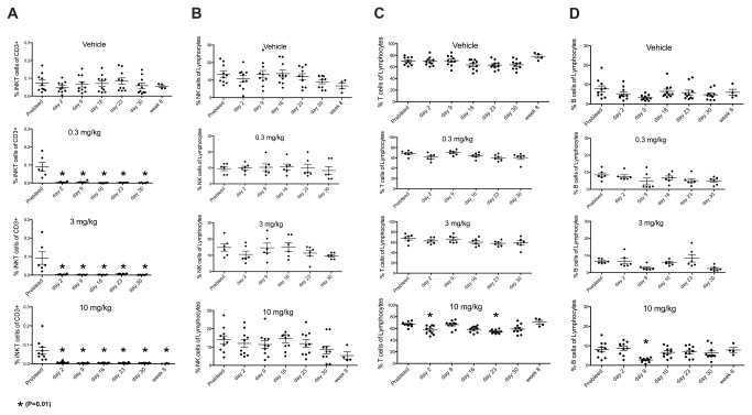
Figure 6. Cynomolgus macaques (n = 5 females + 5 males per group) were dosed and then weekly redosed for 4 weeks (total of 5 doses) with the indicated doses of NKTT120.
Whole blood samples were drawn at indicated timepoints and stained with antibodies against CD3 (SP34.2), CD20 (2H7), TCR Vα24 (C15), CD159a (Z199) and invariant TCR (6B11). Red blood cells were subsequenltly lysed, cells washed and analyzed by flow cytometry. iNKT cells were cells identified as CD3+, Vα24+ and 6B11+ lymphocytes and reported as % of CD3+ cells (A). NK cells were identified as CD3-, CD159a+ lymphocytes and reported as % of lymphocytes (B). T cells were identified as CD3+, CD20- and 6B11- lymphocytes and reported as % of lymphocytes (C). B cells were identified as CD3-, CD20+ lymphocytes and reported as % of lymphocytes (D). One way ANOVA with multiple comparison followed by a non-parametric T test was performed using PRISM software on all the subsets. * indicates significant difference to pre-bleed values (P=0.01).

