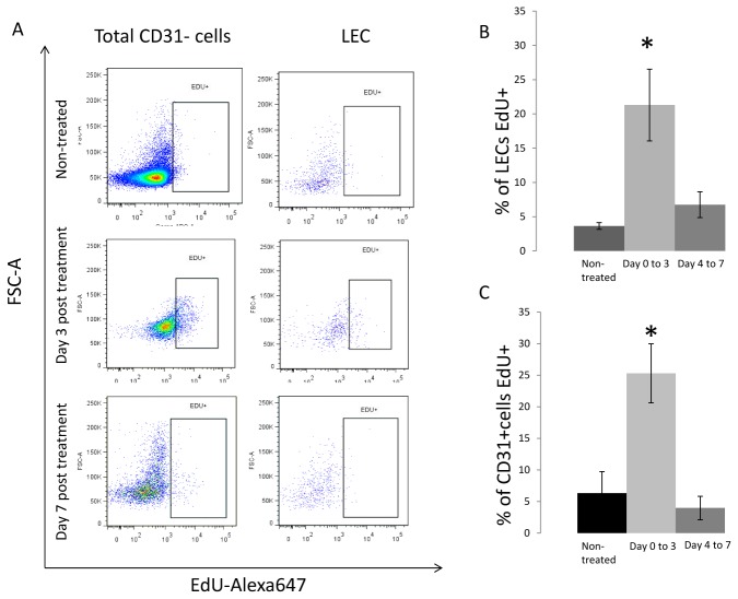
Figure 3. Identification of proliferating LECs.
(A) EdU was injected every 24 hours I.P into mice either without treatment or after CHS. Single cell suspensions were made and viable CD45-/CD31+/ podoplanin+ cells were isolated using FACS. Cells were then fixed with 2% PFA, and stained using click it chemistry with an Alexa647 dye and DAPI. Flow cytometry analysis was performed on the sample to identify DAPI + /EdU + cell population. Sample flow cytometry plot of the FCS-A to EdU fluorescence for all three groups, non-treated, injected from day 0 to day3, and from day 4 to day 7. (B) Quantification of LECs or total CD31+ cells that stained for DAPI + /EdU + in non-treated mice, in mice injected from day 0 to day 3 post CHS, and in mice injected at day 4 to day 7. We observed a significant increase in the number of LECs and total CD31+ cells that intercalated EdU from day 0 to 3, the treated anmials with injection from day 4 to 7, and the non-treated groups (N=3) (p<0.001).

