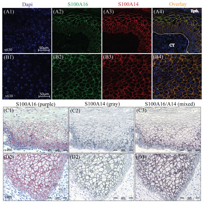
Figure 2. Co-localization of S100A14 and S100A16 in the specimens of OSCC and normal oral mucosa.
Cellular localization of S100A14 and S100A16 was examined by performing DIF and IHC in the sections of formalin fixed and paraffin embedded OSCC and normal oral mucosal tissues. Predominantly membranous localization of S100A16 (green), S100A14 (red) and S100A16/A14 (yellow) was visualized in the epithelial cells of normal mucosa (A1-A4) and OSCC (B1-B4) tissues by DIF (Epth, epithelium; CT, connective tissue). Similarly, with single and double IHC on the serial sections, predominantly membranous expression of S100A16 (purple), S100A14 (gray) and S100A16/A14 (mixed color) was detected in the normal mucosa (C1-C3) and in the OSCC (D1-D3).

